The most common indication for a MRI of the lower extremities is the clarification of the consequences of accidents, unclear pain conditions and inflammatory processes.
MRI can be used to detect inflammation, torn muscle fibres and soft tissue injuries caused by illnesses, accidents or sports injuries, for example.
The examination shows very precisely which structures are affected and how pronounced the changes are.
We then discuss the results with you in detail so that you have clarity about your condition.
Possible examinations
Indications
- Unclear pain
- Hip injuries (bruising, broken bones)
- Injury and inflammation of muscles, tendons and ligaments
- Fatigue fracture of the pelvis and femoral head
- Hip dysplasia
- Detection and characterisation of femoroacetabular impingement
- „Snapping hip“ (coxa saltans)
- Detection of avascular necrosis of the femoral head (HKN)
- Diseases of the femoral head (e.g. osteochondral fracture, osteochondrosis dissecans, transient bone marrow oedema, etc.)
- Assessment of the capsule-ligament structures of the hip joint and the acetabular labrum
- Inflammation of the joint space (arthritis)
- Bursitis (e.g. trochanteric bursitis, iliopsoas bursitis, etc.)
- Inflammatory complications of coxarthrosis
- Perthes' disease (aseptic bone necrosis of the femoral head epiphysis)
- Unclear groin pain (e.g. inflammation of the pubic bone, inguinal hernia, aseptic bone necrosis, etc.)
- Overuse syndromes (e.g. greater trochanter pain syndrome, avulsion fractures, etc.)
- Nerve compression syndromes (e.g. femoral nerve, obturator nerve, sciatic nerve, etc.)
- Suspected metastatic metastases in the bony pelvis
- Tumours
Storage
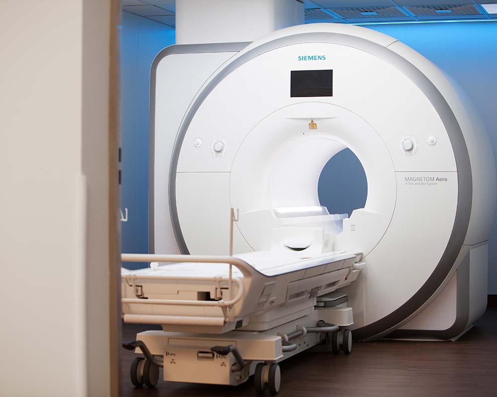
- Comfortably on your back with your head in the centre of the device or with your feet in front and your head outside the tube
Examination time
- approx. 25 minutes
Contrast medium
- This usually takes place MRI examination without the use of a Contrast agent
- However, there are individual questions that cannot be answered with certainty without a contrast agent
- If this is necessary, you will be informed and informed in good time
Indications
- Unclear pain or swelling
- Injuries to the thigh (bruising, broken bones)
- Injury and inflammation of muscles, tendons and ligaments
- Muscle compression syndrome (compartment syndrome)
- Late effects of muscle injuries
- M. Ollier (numerous enchondromas in the metaphyses of the long tubular bones)
- Inflammation of the skeletal muscles (myositis)
- Myositis ossificans
- Tumours
Storage
- Comfortably on your back with your head in the centre of the device or with your feet in front and your head outside the tube
Examination time
- approx. 15 minutes
Contrast medium
- This usually takes place MRI examination without the use of a Contrast agent
- However, there are individual questions that cannot be answered with certainty without a contrast agent
- If this is necessary, you will be informed and informed in good time
Indications
- Unclear pain or swelling
- Degenerative or traumatic meniscus damage (meniscus tear, meniscus degeneration, meniscus cysts)
- Injuries to the knee joint
- Capsule-ligament injuries (e.g. cruciate ligament rupture, rupture of the medial collateral ligament, etc.)
- Injury and inflammation of tendons (e.g. patellar tendon, quadriceps tendon, etc.)
- Traumatic patellar dislocation
- Inflammation of the joint space (arthritis)
- Detection of dysplasia and malpositions of the kneecap (patella)
- Assessment of the articular cartilage
- Osteoarthritis (gonarthrosis)
- Overuse syndromes (e.g. patella tip syndrome, Osgood-Schlatter disease, iliotibial tract friction syndrome, etc.)
- Osteochondrosis dissecans (OD)
- Osteochondral lesions
- M. Ahlbäck (spontaneous osteonecrosis of the knee joint in adults)
- Postoperative conditions (e.g. after partial meniscus resection, after ligament replacement surgery, detection or exclusion of complications such as implant rupture, impingement, hypertrophic scar tissue = cyclops sign, tunnel cysts, Hoffa fibrosis, etc.)
- Pigmented villonodular synovitis (PVNS)
- Bursitis (e.g. prepatellar bursitis, anserine bursitis, Baker's cyst, etc.)
- Detection and characterisation of cysts, ganglia and plicules
- Anomalies (e.g. disc meniscus)
- Tumours
Storage
- Comfortably on your back with your head in the centre of the device or with your feet in front and your head outside the tube
Examination time
- approx. 20 minutes
Contrast medium
- This usually takes place MRI examination without the use of a Contrast agent
- However, there are individual questions that cannot be answered with certainty without a contrast agent
- If this is necessary, you will be informed and informed in good time
Indications
- Unclear pain or swelling
- Injuries to the lower leg (bruising, broken bones)
- Injury and inflammation of muscles, tendons and ligaments
- Shin splints syndrome (shin splints)
- Muscle compression syndrome (compartment syndrome)
- Late effects of muscle injuries
- M. Ollier (numerous enchondromas in the metaphyses of the long tubular bones)
- Inflammation of the skeletal muscles (myositis)
- Myositis ossificans
- Tumours
Storage
- Comfortably on your back with your head in the centre of the device or with your feet in front and your head outside the tube
Examination time
- approx. 15 minutes
Contrast medium
- This usually takes place MRI examination without the use of a Contrast agent
- However, there are individual questions that cannot be answered with certainty without a contrast agent
- If this is necessary, you will be informed and informed in good time
Indications
- Unclear pain or swelling
- Heel pain
- Ligament and syndesmosis injury (following sprain)
- Injuries to the ankle joint (bruises, broken bones)
- Injury and inflammation of muscles, tendons and ligaments
- Osteochondrosis dissecans (OD)
- Osteochondral lesions (e.g. talus)
- Inflammation of the joint space (arthritis)
- Joint instability
- Blockages in the ankle joint (impingement)
- Osteoarthritis
- M. Sever (apophysitis of the calcaneus)
- Sinus tarsi syndrome (inflammatory change in the soft tissue of the sinus tarsi)
- Fatigue fracture (e.g. calcaneus)
- Rheumatoid arthritis
- Tarsal tunnel syndrome (compression syndrome of the tibial nerve)
- Detection and characterisation of lesions / changes in the plantar fascia (e.g. plantar fasciitis, plantar tendon rupture, Ledderhose disease = plantar fibromatosis, etc.)
- Bursitis (e.g. bursitis calcanea inferior)
- Turf toe (injury to the metatarsophalangeal joint of the big toe)
- Tumours
Storage
- Comfortably on your back with your head in the centre of the device or with your feet in front and your head outside the tube
Examination time
- approx. 20 minutes
Contrast medium
- This usually takes place MRI examination without the use of a Contrast agent
- However, there are individual questions that cannot be answered with certainty without a contrast agent
- If this is necessary, you will be informed and informed in good time
Indications
- Unclear pain or swelling
- Degenerative changes (Achilles tendinosis)
- Injury to the Achilles tendon (partial or complete rupture / partial or complete tear)
- Postoperative conditions (detection or exclusion of complications such as rerupture, hypertrophic scar tissue, abscess, etc.)
- Inflammation of the Achilles tendon (tendinitis or paratenonitis)
- Bursitis (bursitis subachillea)
- Anomalies (Haglund's exostosis)
- Tumours
Storage
- Comfortably on your back with your head in the centre of the device or with your feet in front and your head outside the tube
Examination time
- approx. 20 minutes
Contrast medium
- This usually takes place MRI examination without the use of a Contrast agent
- However, there are individual questions that cannot be answered with certainty without a contrast agent
- If this is necessary, you will be informed and informed in good time
Indications
- Unclear pain or swelling
- Injuries to the foot (bruises, broken bones)
- Injury and inflammation of muscles, tendons and ligaments
- Fatigue fracture (mostly ossa metatarsalia II and III/ 2nd and 3rd metatarsal bone)
- Detection and characterisation of changes in the sesamoid bones (inflammation or fracture)
- Osteoarthritis
- Heel spur
- Bursitis (e.g. intermetatarsal bursitis)
- Anlage variants (e.g. Os naviculare accesorium, Os tibiale externum, etc.)
- Baxter's neuropathy (compression syndrome of the inferior calcaneal nerve)
- Morton's neuroma (painful disease caused by compression of the plantar nerves between the heads of the metatarsals)
- Foot deformities (e.g. coalitio calcaneonavicularis, talus verticalis, etc.)
- Diabetic foot
- Rheumatoid arthritis
- Gout
- Tumours
Storage
- Comfortably on your back with your head in the centre of the device or with your feet in front and your head outside the tube
Examination time
- approx. 20 minutes
Contrast medium
- This usually takes place MRI examination without the use of a Contrast agent
- However, there are individual questions that cannot be answered with certainty without a contrast agent
- If this is necessary, you will be informed and informed in good time