The most common indication for a MRI of the upper extremities is the clarification of the consequences of accidents, unclear pain conditions and inflammatory processes.
The examination helps you to better understand pain or restricted movement and to narrow down possible causes.
You receive a detailed visualisation of the affected structures, which facilitates targeted further treatment.
We then explain the results to you in an understandable way so that you know what to do next.
Possible examinations
Indications
- Unclear pain
- Bottleneck syndrome (impingement)
- Rotator cuff rupture
- Labral injuries
- Calcified shoulder (tendinitis calcarea or periarthritis calcarea)
- Inflammation of the shoulder (shoulder arthritis)
- Frozen shoulder (frozen shoulder or capsulitis)
- Bursitis (subacromial bursitis)
- Shoulder injuries (bruises, broken bones)
- Injury and inflammation of muscles, tendons and ligaments
- Dislocation of the joint (luxation)
- Injury or osteoarthritis of the acromioclavicular joint (acromioclavicular joint)
- Osteoarthritis of the shoulder joint (omarthrosis)
- Shoulder instability
- Postoperative conditions (rotator cuff surgery, subacromial decompression, instability surgery, etc.)
- Rheumatoid arthritis
- Nerve compression syndromes
- Tumours
Storage
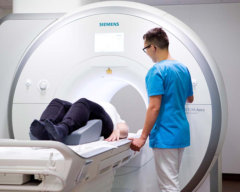
- Comfortable on the back with the head in the centre of the device
Examination time
- approx. 20 minutes
Contrast medium
- This usually takes place MRI examination without the use of a Contrast agent
- However, there are individual questions that cannot be answered with certainty without a contrast agent
- If this is necessary, you will be informed and informed in good time
Indications
- Unclear pain
- Osteolysis syndrome of the clavicle
- Injuries (bruises, broken bones)
- Acromioclavicular joint fusion (ACG fusion)
- Acromioclavicular joint arthrosis (AC joint arthrosis)
- Inflammation of the acromioclavicular joint (AC joint arthritis)
- Anomaly (congenital pseudarthrosis, hypoplasia, aplasia, etc.)
- Tumours
Storage
- Comfortable on the back with the head in the centre of the device
Examination time
- approx. 20 minutes
Contrast medium
- This usually takes place MRI examination without the use of a Contrast agent
- However, there are individual questions that cannot be answered with certainty without a contrast agent
- If this is necessary, you will be informed and informed in good time
Indications
- Unclear pain or swelling
- Injuries to the upper arm (bruising, broken bones)
- Injury and inflammation of muscles, tendons and ligaments
- Muscle compression syndrome (compartment syndrome)
- Late effects of muscle injuries
- M. Ollier (numerous enchondromas in the metaphyses of the long tubular bones)
- Inflammation of the skeletal muscles (myositis)
- Myositis ossificans
- Tumours
Storage
- Comfortable on the back with the head in the centre of the device
Examination time
- approx. 20 minutes
Contrast medium
- This usually takes place MRI examination without the use of a Contrast agent
- However, there are individual questions that cannot be answered with certainty without a contrast agent
- If this is necessary, you will be informed and informed in good time
Indications
- Unclear pain or swelling
- Injuries to the elbow (bruises, broken bones)
- Characterisation of tendon and ligament injuries (e.g. rupture of the distal biceps tendon, ruptures of the triceps tendon, injuries to the collateral ligaments, etc.)
- Injury and inflammation of muscles, tendons and ligaments
- Dislocation of the joint (luxation)
- Tennis elbow or tennis elbow (epicondylitis humeri radialis)
- Golfer's elbow or golfer's elbow (epicondylitis humeri ulnaris)
- Osteochondrosis dissecans (OD)
- M. Panner (aseptic bone necrosis of the humeral capitulum)
- Synovial diseases (e.g. PVNS=pigmented villonodular synovitis, synovial chondromatosis, synovial proliferation of rheumatoid arthritis, etc.)
- Cubital tunnel synrome (sulcus ulnaris syndrome)
- Supinator tunnel syndrome (compression of the posterior interosseous nerve)
- Pronator teres syndrome (compression of the median nerve)
- Bursitis (e.g. bursitis olecrani)
- Rheumatoid arthritis
- Tendon and tendon sheath inflammation (e.g. biceps tendon)
- Tumours
Storage
- Comfortable on your stomach, with the arm to be examined extended forwards; you move your head to the first third of the examination tunnel and can look sideways outwards
Examination time
- approx. 15 minutes
Contrast medium
- This usually takes place MRI examination without the use of a Contrast agent
- However, there are individual questions that cannot be answered with certainty without a contrast agent
- If this is necessary, you will be informed and informed in good time
Indications
- Unclear pain or swelling
- Injuries to the forearm (bruises, broken bones)
- Injury and inflammation of muscles, tendons and ligaments
- Essex-Lopresti injury
- Muscle compression syndrome (compartment syndrome)
- Late effects of muscle injuries
- M. Ollier (numerous enchondromas in the metaphyses of the long tubular bones)
- Inflammation of the skeletal muscles (myositis)
- Myositis ossificans
- Tumours
Storage
- Comfortable on your stomach, with the arm to be examined extended forwards; you move your head to the first third of the examination tunnel and can look sideways outwards
Examination time
- approx. 20 minutes
Contrast medium
- This usually takes place MRI examination without the use of a Contrast agent
- However, there are individual questions that cannot be answered with certainty without a contrast agent
- If this is necessary, you will be informed and informed in good time
Indications
- Unclear pain or swelling
- Wrist injuries (bruises, broken bones)
- Injury and inflammation of muscles, tendons and ligaments
- TFCC lesions
- Ulnolunar impaction syndrome
- Scaphoid fracture and necrosis
- Wrist instabilities
- M. Kienböck (lunate malacia)
- M. Preiser (aseptic bone necrosis of the navicular bone)
- M. Sudeck's disease (complex regional pain syndrome)
- Ganglia
- Osteoarthritis of the carpal bones
- Joint inflammation (arthritis)
- Tendon and tendon sheath inflammation (e.g. tendovaginitis stenosans de Quervain, „intersection syndrome“, tendovaginitis of the extensor carpi ulnaris tendon, etc.)
- Rheumatoid arthritis
- Carpal tunnel syndrome (compression syndrome of the median nerve)
- Syndrome of the Loge de Guyon (compression syndrome of the ulnar nerve)
- Anomaly (e.g. ulna-plus variant, ulna-minus variant, carpal coalition, etc.)
- Tumours
Storage
- Comfortable on your stomach, with the arm to be examined extended forwards; you move your head to the first third of the examination tunnel and can look sideways outwards
Examination time
- approx. 15 minutes
Contrast medium
- This usually takes place MRI examination without the use of a Contrast agent
- However, there are individual questions that cannot be answered with certainty without a contrast agent
- If this is necessary, you will be informed and informed in good time
Indications
- Unclear pain or swelling
- Injuries to the fingers (bruises, broken bones)
- Injury and inflammation of muscles, tendons and ligaments
- Tendonitis and tendon sheath inflammation
- Palmar fibromatosis (Dupuytren's disease)
- Rheumatoid arthritis
- Thumb saddle joint arthrosis (rhizarthrosis)
- Goalkeeper's or skier's thumb (ulnar collateral ligament rupture at the metacarpophalangeal joint of the thumb)
Storage
- Comfortable on your stomach, the hand to be examined extended forwards; you move your head up to the first third of the examination tunnel and can look sideways outwards
Examination time
- approx. 15 minutes
Contrast medium
- This usually takes place MRI examination without the use of a Contrast agent
- However, there are individual questions that cannot be answered with certainty without a contrast agent
- If this is necessary, you will be informed and informed in good time