On this page we have collected and answered the most frequently asked questions about our examinations in everyday practice. Please click on one of the following questions to see the corresponding answer directly. Our aim is to provide you with all the important information about your examination in an easy-to-understand way. If your question is not listed here, you can write us a message at any time or contact our practice team in person. We will be happy to take the time to answer your enquiry.

Frequently asked questions
The abbreviation MRI stands for magnetic resonance imaging, also known as magnetic resonance imaging (MRI) or, colloquially, nuclear spin. It is a frequently used imaging procedure that is used to create precise, high-resolution cross-sectional images of the body. The doctor can use these images to assess organ structures and functions. If the entire body is examined using magnetic resonance imaging, this is referred to as a whole-body MRI. However, only individual body parts or organs can also be examined.
Nuclear magnetic resonance imaging utilises the fact that atomic nuclei rotate around their own axis. This rotation is called nuclear spin and generates a small magnetic field around each nucleus. The hydrogen atoms found throughout the human body also exhibit this nuclear spin. Normally, their axes of rotation point in different directions. However, this changes with magnetic resonance imaging: the MRI machine (magnetic resonance tomograph) is usually a large tube into which the patient is pushed on a couch. The ring-shaped magnetic tunnel generates a strong magnetic field along which the hydrogen atoms in the patient's body are aligned in parallel. The MRI machine then emits short radio wave pulses, which briefly move the hydrogen atoms out of position. The atoms also absorb some energy in the process. After each pulse, they return to the parallel alignment. This process is known as relaxation. The previously absorbed energy, which the hydrogen atoms release again, is recorded. As the different tissues in the body have different water contents, this results in different signals from which the computer calculates the MRI images.
Computed tomography and magnetic resonance imaging are both cross-sectional imaging procedures, i.e. the organ area to be examined is displayed in layers of a few millimetres thickness without overlapping. The main difference lies in the underlying physical principle: CT works with X-rays. MRI converts the magnetic resonance of our body's hydrogen atoms into image information and does not require any radiation at all.
No. So far No harmful effects known. Minimal heating may occur in the tissue. However, there are very low limit values that are always adhered to. We will always provide you with hearing protection (headphones or earplugs) against the loud noises. There are therefore no rules as to how many examinations you can have or at what intervals.
Magnetic resonance imaging does not use ionising radiation, unlike conventional X-ray examinations or computer tomography. You are therefore not exposed to Radiation or radioactivity.
People with pacemakers, insulin pumps, neurostimulators, cochlear implants and defibrillators cannot be examined or can only be examined with the special authorisation of the examining doctor. The examination is critical and must also be considered on a case-by-case basis by your examining doctor if metal splinters are known to be present in the eye socket or facial area, if surgery has been performed on cerebral vessels (e.g. closure of an aneurysm) and if the auditory ossicles in the tympanic cavity have been surgically replaced a long time ago. Please inform us of this when you register or call us in advance. Endoprostheses (knee, hip), vascular operations with the insertion of stents, the vast majority of heart valves, metal wires remaining in the sternum after bypass operations and also dental fillings, bridges etc. are not a problem.
In most cases, appointments are made by telephone on 069-677016350. The appointment service is available on all working days from 7.30 am to 6.00 pm. Outside of these hours, you can use our call-back service or send us an e-mail to info@radiologie-dr-jung.de. About our Online appointment booking you can make an appointment for an examination at our practice at any time.
Yes, you should not change your normal medication. No relevant interactions with the contrast media we use are known to date.
Examinations of the abdominal cavity, brain and blood vessels are often carried out with contrast medium, while examinations of the joints and spine are only occasionally performed. Many MRI examinations are carried out without contrast medium. However, in some cases a contrast agent is indicated for a complete clarification. A thin, flexible plastic tube is inserted into a vein in the arm for the duration of the examination and the contrast agent is injected via a pump. Some tissues, such as muscles and blood vessels, appear in similar shades of grey in the sectional images and are therefore difficult to distinguish. Blood vessels can be better visualised with the help of a contrast agent. Contrast medium is injected into the arm vein and is distributed throughout the body via the bloodstream. It also accumulates more in tumours and metastases, making them clearly visible. A frequently used contrast medium is Gadolinium DOTA. The contrast medium used is not an iodine-containing X-ray contrast medium. It contains chemical compounds of gadolinium or iron. Therefore, if you have had a hypersensitivity to contrast media during an X-ray examination, you can still be examined with MRI/MRI contrast media. These are much better tolerated than X-ray contrast media containing iodine. An overactive thyroid (hyperthyroidism) is no obstacle to the administration of an MRI/MRI contrast agent. Allergy sufferers have a theoretically slightly higher rate of hypersensitivity to MRI/MRI contrast media. The antidotes for allergic reactions are of course available.
Gadolinium is a metal. Taken on its own, gadolinium would be toxic - in contrast media, however, it is firmly bound to a non-toxic carrier substance so that it cannot easily dissolve in the blood. Side effects rarely occur after contrast medium administration: A feeling of warmth after the injection, tingling or skin irritation or temporary discomfort are possible. The symptoms usually subside quickly on their own. Allergies are very rare. Anyone who is prone to allergies should inform the doctors of this in the preliminary consultation. If you notice any unusual symptoms during or after the examination, you should not hesitate to speak to the staff about them.
Contrast agents containing gadolinium are often used to better detect changes and tumours in the brain. Studies have now shown that some of the contrast agents can be deposited in the brain. To date, no harmful effects of gadolinium deposits in the brain are known. However, long-term risks cannot yet be ruled out with certainty. Therefore, as a precaution: since February 2018, the corresponding contrast agents have no longer been used in Germany. Caution also applies to kidney damage: gadolinium compounds can also be deposited in the body, for example in internal organs, the skin or bones. This is particularly the case if the contrast medium is not excreted as quickly in patients with kidney damage and therefore remains in the blood longer than usual. In some patients, changes in the connective tissue of the organs occurred. Patients with kidney damage are therefore only given contrast media if there is no other option.
Compared to many other devices, the magnet opening of the Magnetom Aera is wider at 70 centimetres. Due to the extremely short magnet, many examinations can also be carried out with the patient's head outside the magnet. In addition, the system contains noise-reduced sequences in which the volume is significantly reduced by optimised and intelligent switching of the gradients - while maintaining the same image quality and examination time. Some measurements are even completely noiseless. All of this ensures a relaxed examination atmosphere, which particularly benefits patients with claustrophobia. The newly designed casing of the device is also equipped with MoodLight illumination, which creates a friendly and colourful environment in the examination room.
If the foot, lower leg, knee or hip are being examined, your upper body remains outside the tunnel. The head and upper body can also remain outside the tunnel during an examination of the lumbar spine. A bell is placed in your hand and you are in contact with Beyond Imaging staff at all times via an intercom system. A camera at the head end provides additional safety. Your companion is welcome to sit with you during the examination. The Magnetic tunnel is bright, well ventilated and has friendly, rounded contours. The room can be illuminated in different colours according to your wishes. We will make your examination as short as possible.
In general, we recommend that you wear comfortable clothing that you feel comfortable in for the examination. Please note that any kind of metal can negatively affect the quality of the MRI images. For examinations in the chest, shoulder, head and pelvic area as well as for examinations of the spine, you should not wear any clothing or underwear with metal hooks or metal fasteners or take them off in the changing room before the examination. All metal objects such as hair clips, earrings, jewellery and piercings must be removed in the changing room before the examination. Trousers with metal buttons, zips, press studs or belts can also have a negative impact on image quality and should be removed before the examination. If required, we can provide you with metal-free clothing for the duration of the examination in exceptional cases. Furthermore, make-up may occasionally interfere with examinations in the head and neck area (some products contain metal particles). Please inform our staff before the examination if you are wearing permanent make-up.
The duration of the examination varies depending on the question being asked and the region being examined. For joint examinations it is usually 15 to 20 minutes, but for examinations of the abdominal cavity it can also be 30 to 40 minutes.
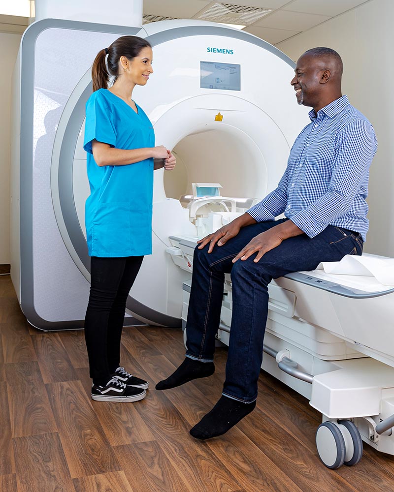
Firstly, the signal-receiving coil is placed around the body region to be examined in the preparation room. You will then be moved from the preparation room - already lying on the MRI/MRI table - to the centre of the cylindrical magnet („tunnel“), because only there is the homogeneous magnetic field that is essential for the examination. The MRI/MRI has a wide diameter of 70 cm and is open at both ends. The inside of the tunnel is equipped with air conditioning and lighting, both of which can be individually adjusted to different levels. During an examination of the knee joint, for example, your knee is in a coil in the centre of the tunnel and your upper body/head is outside the „tube“.
The examination is controlled from an operator station outside the MRI/MRI room. There is voice and visual contact between Beyond Imaging staff and you in the examination room. As the examination is loud for measurement reasons, you will be given headphones (with music if required) or earplugs. You will also be given a bell with which you can make yourself heard at any time during the examination. During the measurements you should relax and please do not move! Otherwise the images will be blurred as the signals emitted by your body cannot be clearly assigned. They may then not be usable and the measurement will have to be repeated. For some examinations it is necessary to use a contrast agent. This type of contrast agent was specially developed for magnetic resonance imaging and is very well tolerated. It increases the diagnostic value of magnetic resonance imaging and differentiates tissue types even more clearly.
After approximately four to six measurements, each lasting one to two minutes, sometimes a little longer, the examination is finished. You will be taken back to the preparation room and can get dressed again.
Our 1.5 Tesla MR device has an extremely short and very wide tube with extremely good image quality. This allows us to offer patients who suffer from claustrophobia an anxiety-free examination in most cases. If necessary, an anxiolytic or sedative medication can be administered before the examination.
If you are under Claustrophobia please talk to us about this. In many cases, examinations can be carried out in such a way that the head remains outside the „tube“. Many people are also reassured if they can take a look at the MRI machine the day before the examination, for example, in order to prepare themselves for the situation. If you suffer from claustrophobia, we can give you a sedative before the examination. We will give you this sedative as a drink, which we have had very good experience with. It is necessary for you to arrive at our practice 30 minutes before your appointment. It is important that you come to the examination with an accompanying person and are taken home afterwards. Once you have been given a sedative, you are not allowed to actively participate in road traffic, operate machinery or climb ladders for 24 hours, i.e. you are not allowed to drive yourself!
Please let us know already at the Appointment allocation if a tranquilliser is required.
There is nothing to be said against the presence of an accompanying person during the examination. Patients who suffer from claustrophobia in particular often benefit from the presence of a familiar person. Of course, all persons present in the MRI are also subject to the safety regulations applicable to patients (metal, pacemakers, etc.).
The Pictures will be given to you after the examination in the form of a CD-ROM. You will also receive personalised access data with which the images from the MRI examination can be called up online. We will send the written findings to the referring doctor within 24 hours by fax, e-mail or post. In medically urgent cases, the written findings will be sent immediately or the referring doctor will be informed by telephone.
There are no known harmful effects for the unborn child, but as a precautionary measure in the first third (trimester) of pregnancy Pregnancy MRI is not performed in the early stages of pregnancy (with the exception of a vital indication for the mother). An MRI is then possible again in late pregnancy.
At most, minimal amounts of the contrast agent could pass into the breast milk and an infant cannot absorb it from the intestine. However, as a purely precautionary measure, we recommend expressing and discarding breast milk for 24 hours after the examination.
Yes, in many cases magnetic resonance imaging is the method of first choice in the diagnosis of paediatric diseases. As the paediatric body is particularly sensitive to radiation, a Radiation exposure by X-ray and CT should be avoided as far as possible. Underage patients must be accompanied by a parent or legal guardian, as minors are not usually able to give their consent by signature after being informed about the planned examination.
Alternatively, young people can bring a written declaration of consent from a parent or legal guardian, but this must be confirmed in advance. Arrangement by telephone and, if necessary, clarification. Please contact us if you have any questions.
Only for special MRT examinations, e.g. the visualisation of the bile ducts (MRCP) or the gastrointestinal tract (MDP, Sellink), is a absolute sobriety required, so that good visualisation is possible. In this case, you must not eat or drink anything from 10 p.m. on the previous day. For all other MRI examinations, you can eat normally before and after the examination.
MRI imaging is associated with Tattoos possible. The tattoo inks used in Germany since the 1990s generally contain few to no magnetic ingredients and are suitable for MRI.
The localisation and size of the tattoo also determine whether an MRI examination is possible and useful. Tattoos in the area of the body part to be examined can interfere with the imaging and result in a lower resolution of the MRI image. The same effects can also be seen with particularly large tattoos in the area of the body to be examined.
As a general rule, all metal jewellery should be removed before an MRI examination if possible. Piercings can be made of different metals, which are magnetic to varying degrees. The metals iron, cobalt and nickel in particular can be moved by the magnetic field and heat up. For this reason, an MRI examination with piercings made of these materials is generally ruled out for safety reasons.
Piercings made of titanium, implantanium or polytetrafluoroethylene (PTFE) pose no risk to the patient in an MRI scan. These metals are only weakly magnetic or non-magnetic and can therefore neither be attracted nor heated by the magnetic field. For this reason, it is possible to perform MRI imaging with these piercings as long as they are not located in the body section to be examined. They can then cover the underlying structures and affect the image quality. If the patient does not know the exact composition of the piercing materials, a piercing should always be removed for safety reasons. A plastic piercing is made of plastic. Plastic is not magnetic and therefore does not react to the strong magnetic field in the MRI. It is therefore not necessary to remove a plastic piercing before the examination. The plastic piercing can still be worn even if imaging is carried out in the area of the plastic piercing. Many piercing studios offer to replace metal piercings with plastic piercings for an MRI examination.
Yes, these implants are MR-compatible. However, the image quality may be limited in the corresponding areas.
If you are to have an examination with contrast medium injection, we need the so-called creatinine value. This is a laboratory value that your general practitioner or specialist can determine for you. Ideally, the value should not be older than 4 weeks. However, if you have no previous kidney disease, older values may also be sufficient. If in doubt, you should ask when you make your appointment. If available, you should bring along documents relating to allergies (allergy passport) and implants.
You should arrive at the registration desk approx. 20 minutes before the start of the examination. No special preparation is necessary for most examinations. In particular, you do not need to be fasting. However, if you require an examination of the Abdomen (upper abdominal organs, liver, gall bladder, pancreas, etc.) is planned, you should only consume a small amount of food and keep a certain distance before the examination. Please let us know if you are wearing any items containing metal, in particular
- Vascular supports such as stents or vascular clips
- an artificial heart valve
- an insulin pump
- Metal splinters
- Tattoos
In principle, it always makes sense for us to have precise information about your (pre-)illnesses. Comparative images are an advantage when it comes to assessing the course of a disease or detecting subtle changes. Therefore, please bring the images in your possession or archived by your doctor to the examination.
If you have been examined by us recently, you can assume that your images are stored in our archive system. It is then not necessary to bring the previous images with you.
We work with the most modern MRI devices available on the market. Our MRTs have a very Short tube, which opens wide at both ends, with a diameter of 70 cm. A bell is placed in your hand and you are in contact with Beyond Imaging staff at all times via an intercom system. A camera at the head end provides additional safety. Your companion is welcome to sit with you during the examination. For many examinations, your upper body is outside the tube of the magnet. The magnet tunnel is bright, well ventilated and has friendly, round contours. The room can be illuminated in different colours according to your wishes. We will make your examination as short as possible.
According to current scientific knowledge, magnetic resonance imaging does not have any harmful effects or side effects. Magnetic resonance imaging does not use X-rays, so repeated examinations are not harmful.